Open Access
Peer Reviewed
ARTICLES
2613 Viewed1120 Downloaded
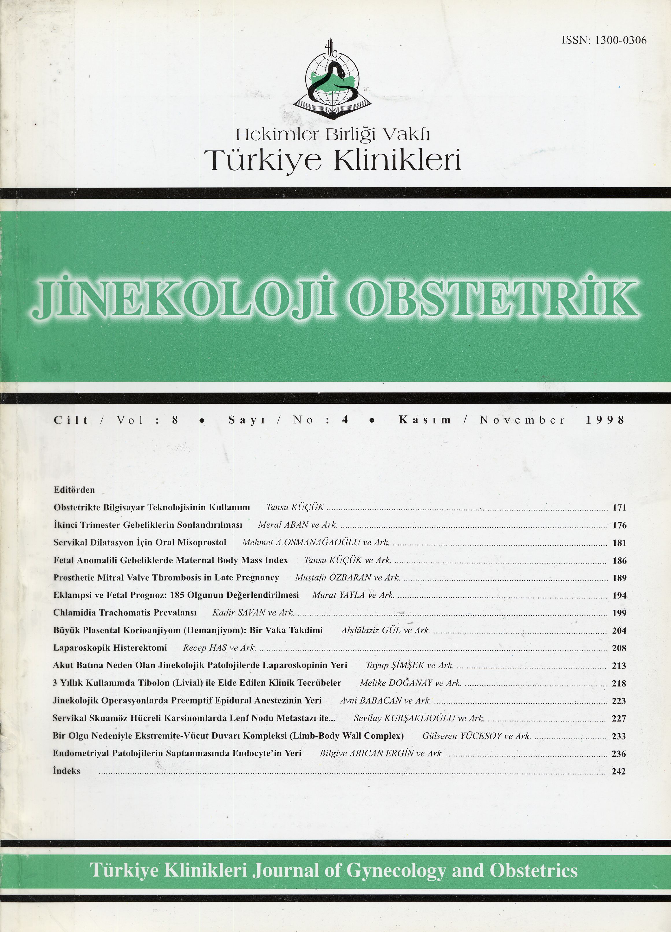
Large Chorioangioma (Hemangioma) Of The Placenta: A Case Report
Büyük Plasental Korioanjiyom (Hemanjiyom):Bir Vaka Takdimi
Turkiye Klinikleri J Gynecol Obst. 1998;8(4):204-7
Article Language: TR
Copyright Ⓒ 2020 by Türkiye Klinikleri. This is an open access article under the CC BY-NC-ND license (http://creativecommons.org/licenses/by-nc-nd/4.0/)
ÖZET
Amaç: Kliniğimizde teşhis edilen büyük plasental hemanjiyom olgusunu sunmak, tanı ve komplikasyonlarını vurgulamak. Çalışmanın yapıldığı yer: Yüzüncü Yıl Üniversitesi Tıp Fakültesi Kadın Hastalıkları ve Doğum ABD Van. Materyal ve Metod: Bu yazıda 23 yaşında primigravid, normal spontan doğumun gerçekleştirildiği, makroskopik ve histopatolojik olarak plasental hemanjiyom tanısı konulan bir vaka sunuldu. Bulgular: Son mens tarihine göre 37 haftalık gebe iken yapılmış olan obsterik ultrasonogarfisinde; plasenta korpus posteriorda ve grade I matürasyonda, baş pelvisde, amnios mayii yeterli olarak tesbit edildi. Normal olmayan görünümü nedeniyle plasenta patolojiye gönderildi. Mikroskopik olarak çoğunluğu küçük çapta olmak üzere değişik çapta lümenleri tek sıra endotelle döşeli ve eritrositler içeren çok sayıda damarın oluşturduğu tümöral yapı görüldü. Bu damarları gevşek bir stroma destekliyordu. Bu bulgularla kapiller hemanjiyom tanısı konuldu. Olgumuzda; Maternal, fetal ve neonatal herhangi bir komplikasyon gözlenmedi. Sonuç: Plasental Korioanjiyom (hemanjiyom) tümör görünümünde bir damar hamartom olup nadir görülmektedir. Maternal, fetal veya neonatal komplikasyonlara yol açması ve özellikle preeklampsi ve eklampsi ile birlikte seyredebilmesi plasental hemanjiyomun önemini artırmaktadır.
Amaç: Kliniğimizde teşhis edilen büyük plasental hemanjiyom olgusunu sunmak, tanı ve komplikasyonlarını vurgulamak. Çalışmanın yapıldığı yer: Yüzüncü Yıl Üniversitesi Tıp Fakültesi Kadın Hastalıkları ve Doğum ABD Van. Materyal ve Metod: Bu yazıda 23 yaşında primigravid, normal spontan doğumun gerçekleştirildiği, makroskopik ve histopatolojik olarak plasental hemanjiyom tanısı konulan bir vaka sunuldu. Bulgular: Son mens tarihine göre 37 haftalık gebe iken yapılmış olan obsterik ultrasonogarfisinde; plasenta korpus posteriorda ve grade I matürasyonda, baş pelvisde, amnios mayii yeterli olarak tesbit edildi. Normal olmayan görünümü nedeniyle plasenta patolojiye gönderildi. Mikroskopik olarak çoğunluğu küçük çapta olmak üzere değişik çapta lümenleri tek sıra endotelle döşeli ve eritrositler içeren çok sayıda damarın oluşturduğu tümöral yapı görüldü. Bu damarları gevşek bir stroma destekliyordu. Bu bulgularla kapiller hemanjiyom tanısı konuldu. Olgumuzda; Maternal, fetal ve neonatal herhangi bir komplikasyon gözlenmedi. Sonuç: Plasental Korioanjiyom (hemanjiyom) tümör görünümünde bir damar hamartom olup nadir görülmektedir. Maternal, fetal veya neonatal komplikasyonlara yol açması ve özellikle preeklampsi ve eklampsi ile birlikte seyredebilmesi plasental hemanjiyomun önemini artırmaktadır.
ANAHTAR KELİMELER: Plasental Korioanjiyom; Feto-Maternal Komplikasyonlar
ABSTRACT
Objective: To present ?a large placental Chorioangioma case that was diagnosed and treated in our department and, to emphasize diagnosis and complications of such cases. Institution: Yüzüncü Yıl Üniversity Medical Faculty, Department of Obstetrics and Gynecology, VAN. Materials and Methods: In this paper, a 23 year old primigravid case that had a normal spontaneus birth, and that was macroscopically and histopatologically diagnosed as plasental hemangioma is presented. Findins: At the end of 37 weeks of gestation, Obstetrics Ultrasonografic results were observed as follows: placenta was located at corpus posterior, the head of infant was in the pelvis and, Amnion Fluid index was normal. After delivery, the placenta was sent for histopathological and macroscopcal examining because of abnormal size. Microscopically, a tumoral structure which was origined with a number of vessels of different diameter, and with endothelial layer as a single line cell and which contained erythrocyte(s) was seen. These vessels were fortified with loose stroma. These findings were evaluated as Capiller Hemangiyoma diagnosis. Because of this case which has not maternal fetal and neonatal complication, the clinical and the histopathological aspects of plasental hemongiom was discussed. Conclusion: Placental Chorioangioma (hamengioma) is a vascular hemartom appears as a tumor. It is a rare seen hemangioma. Causing maternal fetal or neonatal complications and encompaining preaclampsia and eclampsia increases the importance of plasental hemangioma.
Objective: To present ?a large placental Chorioangioma case that was diagnosed and treated in our department and, to emphasize diagnosis and complications of such cases. Institution: Yüzüncü Yıl Üniversity Medical Faculty, Department of Obstetrics and Gynecology, VAN. Materials and Methods: In this paper, a 23 year old primigravid case that had a normal spontaneus birth, and that was macroscopically and histopatologically diagnosed as plasental hemangioma is presented. Findins: At the end of 37 weeks of gestation, Obstetrics Ultrasonografic results were observed as follows: placenta was located at corpus posterior, the head of infant was in the pelvis and, Amnion Fluid index was normal. After delivery, the placenta was sent for histopathological and macroscopcal examining because of abnormal size. Microscopically, a tumoral structure which was origined with a number of vessels of different diameter, and with endothelial layer as a single line cell and which contained erythrocyte(s) was seen. These vessels were fortified with loose stroma. These findings were evaluated as Capiller Hemangiyoma diagnosis. Because of this case which has not maternal fetal and neonatal complication, the clinical and the histopathological aspects of plasental hemongiom was discussed. Conclusion: Placental Chorioangioma (hamengioma) is a vascular hemartom appears as a tumor. It is a rare seen hemangioma. Causing maternal fetal or neonatal complications and encompaining preaclampsia and eclampsia increases the importance of plasental hemangioma.
MENU
POPULAR ARTICLES
MOST DOWNLOADED ARTICLES


This journal is licensed under a Creative Commons Attribution-NonCommercial-NoDerivatives 4.0 International License.


