Open Access
Peer Reviewed
ARTICLES
2507 Viewed915 Downloaded
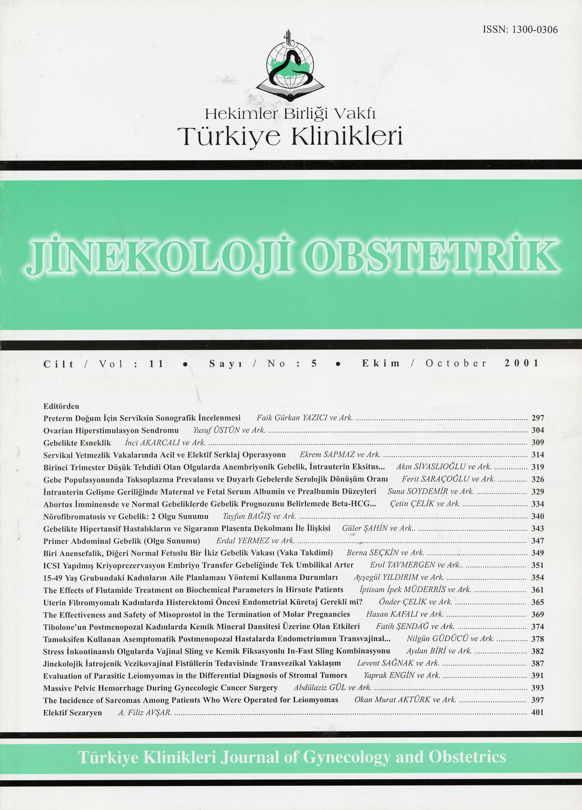
Evaluation Of The Endometrium By Transvaginal Sonography,saline Infusion Sonography, Hysteroscopy And Endometrial Curettage In Asymptomatic Postmenopausal Patients Using Tamoxifen
Tamoksifen Kullanan Asemptomatik Postmenopozal Hastalarda Endometriumun Transvajinal Ultrasonografi, Salin İnfüzyon Sonografi, Histeroskopi ve Endometrial Küretaj İle Değerlendirilmesi
Turkiye Klinikleri J Gynecol Obst. 2001;11(5):378-81
Article Language: TR
Copyright Ⓒ 2020 by Türkiye Klinikleri. This is an open access article under the CC BY-NC-ND license (http://creativecommons.org/licenses/by-nc-nd/4.0/)
ÖZET
Amaç: Meme kanserli postmenopozal hastaların hormonal tedavisinde kullanılan tamoksifen, bir endometrial karsinojen olarak sınıflandırılmıştır. Ancak bu hasta grubunda endometrial patolojilerin tespit edilebilmesi için nasıl bir takip yapılması gerektiği konusunda fikir birliğine varılamamıştır. Bu çalışmada hastanemiz onkoloji kliniğinde tamoksifen ile tedavi edilen postmenopozal hastalara ait endometrial patolojileri değerlendirdik. Çalışmanın Yapıldığı Yer: Dr.Lütfi Kırdar Kartal Eğitim ve Araştırma Hastanesi. Materyel ve Metod: Bir yıldan daha uzun süredir tamoksifen kullanan asemptomatik postmenopozal hastalar transvajinal ultrasonografi (TVUSG) ve salin infüzyon sonografi (SİS) ile değerlendirildi ve endometrial kalınlığın 5mm ve üzerinde ölçüldüğü hastalara histeroskopi ve endometrial küretaj yapıldı. Bulgular: Tamoksifen kullanan 14 postmenopozal hastanın katıldığı çalışmada yedi hastada endometrial kalınlık TVUSG ile 5mmnin üzerinde ölçüldü. Ondört hastaya çekilen SİSlerde TVUSGde endometrial kalınlığın 5mmnin üzerinde ölçüldüğü yedi hastada bir sübmüköz myom, dört endometrial polip bulundu, bir hastada SİS ile kaçırılan endometrial polip histeroskopi ile yakalandı. Bir hastaya ise servikal stenoz nedeniyle SİS çekilemedi. TVUSG ile endometrial kalınlığın 5mmnin altında ölçüldüğü hastalarda SİS ile ek bir patoloji saptanmadı. Histeroskopi ile endometrial polip tespit edilen beş hastanın hepsinde endometrial küretaj sonucu negatif geldi. Sonuçlar: Tamoksifen kullanan hastalarda endometrial patolojilerde artış sözkonusudur, ancak bu hastalarda atrofik zeminde gelişen fokal hiperplastik lezyonlar endometrial küretaj ile kaçırılabilir. Bu nedenle bu hastalarda kullanılabilecek tarama yöntemlerini araştıran çalışmalarda altın standard olarak histeroskopiyi almak gerekir.
Amaç: Meme kanserli postmenopozal hastaların hormonal tedavisinde kullanılan tamoksifen, bir endometrial karsinojen olarak sınıflandırılmıştır. Ancak bu hasta grubunda endometrial patolojilerin tespit edilebilmesi için nasıl bir takip yapılması gerektiği konusunda fikir birliğine varılamamıştır. Bu çalışmada hastanemiz onkoloji kliniğinde tamoksifen ile tedavi edilen postmenopozal hastalara ait endometrial patolojileri değerlendirdik. Çalışmanın Yapıldığı Yer: Dr.Lütfi Kırdar Kartal Eğitim ve Araştırma Hastanesi. Materyel ve Metod: Bir yıldan daha uzun süredir tamoksifen kullanan asemptomatik postmenopozal hastalar transvajinal ultrasonografi (TVUSG) ve salin infüzyon sonografi (SİS) ile değerlendirildi ve endometrial kalınlığın 5mm ve üzerinde ölçüldüğü hastalara histeroskopi ve endometrial küretaj yapıldı. Bulgular: Tamoksifen kullanan 14 postmenopozal hastanın katıldığı çalışmada yedi hastada endometrial kalınlık TVUSG ile 5mmnin üzerinde ölçüldü. Ondört hastaya çekilen SİSlerde TVUSGde endometrial kalınlığın 5mmnin üzerinde ölçüldüğü yedi hastada bir sübmüköz myom, dört endometrial polip bulundu, bir hastada SİS ile kaçırılan endometrial polip histeroskopi ile yakalandı. Bir hastaya ise servikal stenoz nedeniyle SİS çekilemedi. TVUSG ile endometrial kalınlığın 5mmnin altında ölçüldüğü hastalarda SİS ile ek bir patoloji saptanmadı. Histeroskopi ile endometrial polip tespit edilen beş hastanın hepsinde endometrial küretaj sonucu negatif geldi. Sonuçlar: Tamoksifen kullanan hastalarda endometrial patolojilerde artış sözkonusudur, ancak bu hastalarda atrofik zeminde gelişen fokal hiperplastik lezyonlar endometrial küretaj ile kaçırılabilir. Bu nedenle bu hastalarda kullanılabilecek tarama yöntemlerini araştıran çalışmalarda altın standard olarak histeroskopiyi almak gerekir.
ANAHTAR KELİMELER: Tamoksifen, Endometrium, Salin infüzyon sonografi, Histeroskopi
ABSTRACT
bjective: Tamoxifen is a drug used in the hormonal treatment of postmenopausal patients and has recently been classified as an endometrial carcinogen. Many techniques have been suggested for screening patients on tamoxifen for the development of endometrial abnormalities, but a concensus has not been reached yet. Institution: Dr. Lütfi Kırdar Kartal Education and Research Hospital. Material and Method: Asymptomatic postmenopausal patients receiving tamoxifen for more than one year were screened by transvaginal ultrasonography (TVUSG) and saline infusion sonography initially. Patients with an endometrial line measured as more than or equal to 5mm at TVUSG had hysteroscopy and than endometrial curettage. Results: 14 asymptomatic postmenopausal patients receiving tamoxifen participated the study and seven of them had an endometrial line more than or equal to 5mm at TVUSG. These 14 patients are also screened by SIS and in seven patients that had an endometrial line more than or equal to 5mm at TVUSG, 4 endometrial polyps and 1 submucous myoma have been detected. One polyp that has been missed by SIS was diagnosed at hysteroscopy. One patient was unable to undergo SIS due to cervical stenosis.In patients that had an endometrial line less than 5mm, no further pathology has been detected by SIS.All of the patients diagnosed to have endometrial polyp at hysteroscopy had negatif endometrial curettage results. Conclusion: Endometrial pathologies are increased in postmenopausal patients using tamoxifen, but focal hyperplastic lesions developing at an atrofic background can be easily missed by endometrial curettage. Therefore studies designed to find out the screning methods for the development of endometrial pathologies should use hysteroscopy as the gold standard.
bjective: Tamoxifen is a drug used in the hormonal treatment of postmenopausal patients and has recently been classified as an endometrial carcinogen. Many techniques have been suggested for screening patients on tamoxifen for the development of endometrial abnormalities, but a concensus has not been reached yet. Institution: Dr. Lütfi Kırdar Kartal Education and Research Hospital. Material and Method: Asymptomatic postmenopausal patients receiving tamoxifen for more than one year were screened by transvaginal ultrasonography (TVUSG) and saline infusion sonography initially. Patients with an endometrial line measured as more than or equal to 5mm at TVUSG had hysteroscopy and than endometrial curettage. Results: 14 asymptomatic postmenopausal patients receiving tamoxifen participated the study and seven of them had an endometrial line more than or equal to 5mm at TVUSG. These 14 patients are also screened by SIS and in seven patients that had an endometrial line more than or equal to 5mm at TVUSG, 4 endometrial polyps and 1 submucous myoma have been detected. One polyp that has been missed by SIS was diagnosed at hysteroscopy. One patient was unable to undergo SIS due to cervical stenosis.In patients that had an endometrial line less than 5mm, no further pathology has been detected by SIS.All of the patients diagnosed to have endometrial polyp at hysteroscopy had negatif endometrial curettage results. Conclusion: Endometrial pathologies are increased in postmenopausal patients using tamoxifen, but focal hyperplastic lesions developing at an atrofic background can be easily missed by endometrial curettage. Therefore studies designed to find out the screning methods for the development of endometrial pathologies should use hysteroscopy as the gold standard.
MENU
POPULAR ARTICLES
MOST DOWNLOADED ARTICLES


This journal is licensed under a Creative Commons Attribution-NonCommercial-NoDerivatives 4.0 International License.


